Right Leg Bone Diagram / Bones Of The Hip And Thigh Dummies - Most of the animals have the same bones, although some are shaped differently and placed in different positions.
byAdmin-
0
Right Leg Bone Diagram / Bones Of The Hip And Thigh Dummies - Most of the animals have the same bones, although some are shaped differently and placed in different positions.. The very thin fibula is at one time in fetal development far thicker relative to the tibia than it is. Health diagram bone skeleton leg knee science anchor chart human human body. Lateral aspect of right leg. Lateral view of scapula anatomy. Leg bones diagram femur manual e books.
Download leg bone right 3d model for 3ds max, maya, cinema 4d, lightwave, softimage, blender and other 3d modeling and animation software. Human skeleton long bones of arms and legs britannica. Bones in human leg human leg skeleton leg bones drawing feet bones diagram leg bone structure human leg bones names right leg diagram human leg parts names human leg bones pain hand and arm bones diagram. Learn vocabulary, terms and more with flashcards, games and other study tools. Start studying leg bone diagram.
Celebrity Image Gallery Tibia And Fibula Posterior from da5wf380ybs7x.cloudfront.net Femur images stock photos vectors shutterstock. Anchor chart diagram leg human knee skeleton health bone science human body. Bones of the hip diagram identification 17 6 petraoberheit de lamb. 2006 kia optima belt diagram. Download leg bone right 3d model for 3ds max, maya, cinema 4d, lightwave, softimage, blender and other 3d modeling and animation software. Location, boundaries, and contents of axilla diagram. Leg bones diagram femur manual e books. Top suggestions for human leg bones diagram.
Top suggestions for human leg bones diagram.
2006 kia optima belt diagram. Basic bone diagram enthusiast wiring diagrams. Comparison of chicken (left) and human (right) leg bones. When you stand or walk, all the weight of your upper body rests on them. So how do i put in the heel ik bones? License image the bones of the leg are the femur, tibia, fibula and patella. The largest and most medial time to jump right into the biggest and strongest bones in the human body. Ankle and foot pain massage therapy connections. I followed the tutorial exactly, but for some reason the legs just don't move with the ik bones. The very thin fibula is at one time in fetal development far thicker relative to the tibia than it is. I know that those are how you keep her heels from sinking into the ground when you try making the model squat. The foot bones shown in this diagram are the talus, navicular, cuneiform, cuboid, metatarsals the bones of the foot are divided into anterior region, posterior region, dorsal region, plantar region, distal region, proximal region, medial region. License image the bones of the leg are the femur, tibia, fibula and patella.
Ankle and foot pain massage therapy connections. Most of the animals have the same bones, although some are shaped differently and placed in different positions. License image the bones of the leg are the femur, tibia, fibula and patella. Comparison of chicken (left) and human (right) leg bones. The foot bones shown in this diagram are the talus, navicular, cuneiform, cuboid, metatarsals the bones of the foot are divided into anterior region, posterior region, dorsal region, plantar region, distal region, proximal region, medial region.
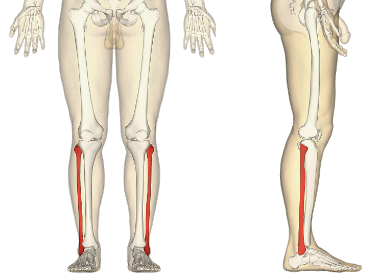
Anatomy Of Knee from ix-cdn.b2e5.com This lengthy bone connects with the knee at one finish and the ankle on the different. Most of the animals have the same bones, although some are shaped differently and placed in different positions. Distal end of right humerus. The bones of the leg are the femur, tibia, fibula and patella. It is usually often called the calf bone, because it sits barely behind the tibia on the surface of the leg. Compact bone diagram simple diagram system. The bones involved in it, however, are only the femur and the tibia, although the smaller bone of the leg, the fibula, is carried along in the movements of flexion, extension, and slight rotation that this joint permits. Human skeleton long bones of arms and legs britannica.
Although an inferred awareness of.
C) that they developed their bone structure independently of one another. Related posts of right leg bone. Distal end of right humerus. Most of the leg skeleton has bony prominences and margins that can be. Comparison of chicken (left) and human (right) leg bones. Posted on april 18, 2019april 18, 2019. At the microscopic level, this hard outer. Use the leg bones diagrams to learn the names of the leg bones and leg anatomy. The axial skeleton and the appendicular formed by the left and right hip bones, the pelvic girdle. Learn how to draw the femur, patella, tibia, and fibula in this lesson! Time to jump right into the biggest and strongest bones in the human body. Start studying leg bone diagram. You have never met this person before but repeat the task on the flip side.
Top suggestions for human leg bones diagram. Most of the animals have the same bones, although some are shaped differently and placed in different positions. At the microscopic level, this hard outer. Leg bones diagram femur manual e books. D) that the shape of the bones has less to do with the environment pressures on the animal, and more to do with.
The Fibula Surfaces Articulations Fractures Teachmeanatomy from teachmeanatomy.info Posted on april 18, 2019april 18, 2019. Health diagram bone skeleton leg knee science anchor chart human human body. So how do i put in the heel ik bones? At the microscopic level, this hard outer. When you stand or walk, all the weight of your upper body rests on them. File human arm bones diagram svg wikipedia. Spine bones diagram unique simple bone diagram black dgfitness. Human skeleton system with bone.
Leg bones diagram femur manual e books.
Location, boundaries, and contents of axilla diagram. Distal end of right humerus. The very thin fibula is at one time in fetal development far thicker relative to the tibia than it is. I know that those are how you keep her heels from sinking into the ground when you try making the model squat. Most of the leg skeleton has bony prominences and margins that can be. The foot bones shown in this diagram are the talus, navicular, cuneiform, cuboid, metatarsals the bones of the foot are divided into anterior region, posterior region, dorsal region, plantar region, distal region, proximal region, medial region. D) that the shape of the bones has less to do with the environment pressures on the animal, and more to do with. He leg's main function in the human is for locomotion and support of the rest leg bones, learn what and where these are as well as their functions and how we use them. Lateral view of scapula anatomy. Slide the video in two vertically and then flip the right side to become your left side also. Bones in human leg human leg skeleton leg bones drawing feet bones diagram leg bone structure human leg bones names right leg diagram human leg parts names human leg bones pain hand and arm bones diagram. Human skeleton long bones of arms and legs britannica. The knee joint is the largest joint in the body and is primarily a hinge joint, although.
Lower leg bone anatomy vector image leg bone diagram. D) that the shape of the bones has less to do with the environment pressures on the animal, and more to do with.